 Click to enlarge
Meiosis Model K20110
Ask About This ProductDescription
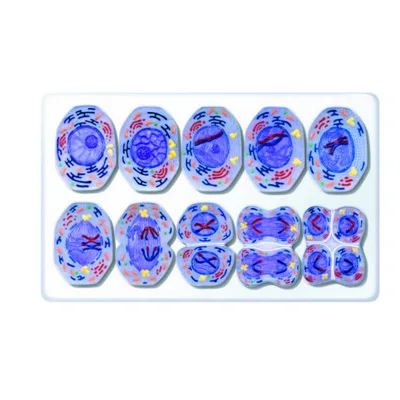
With the meiotic division cell model series, it is possible
to study 10 separate stages of a mammalian cell.
The model is produced enlarged about 10,000
times relative to actual size.
1. Interphase (G1 phase)
2. Prophase I (Leptotene)
3. Prophase I (Zygotene and Pachytene)
4. Prophase I (Diplotene)
5. Prophase I (Diakinesis)
6. Metaphase I
7. Anaphase I
8. Telophase I, Cytokinesis I, Interkinesis,
Prophase II and Metaphase II
9. Anaphase II
10. Telophase II and Cytokinesis II
Coloured on the basis of microscopy technique,
the three-dimensional model makes cell division
easier to understand.
Cell organelles are shown on the lower part
of the models. Each model has a magnet on the back,
so it can easily be displayed in the classroom
on a magnetic board.
All the models
come with a 40 x 60 cm board.
For use in lessons, brochures and
detailed information notes are supplied
with the mitosis model.
Model Dimensions: 60x40x6 cm 1.7 kgDelivery and Installation
Before shipment, our products are inspected and packed in special impact-resistant packaging, and are delivered safely to every province of Türkiye.
Installation: In laboratory and classroom projects, installation and commissioning after delivery are carried out by our expert team.
Training and support: User training and after-sales technical support are provided for experiment sets and simulators.